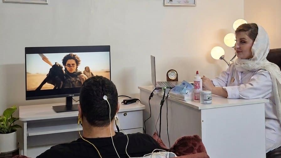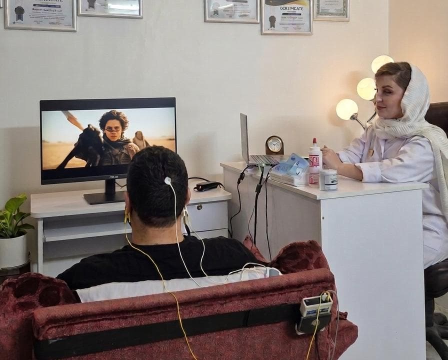
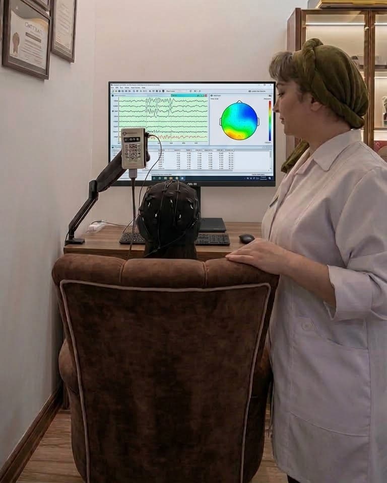
When you hear the term “brain map,” it often refers to QEEG, or quantitative electroencephalography. The name sounds technical, but the idea is straightforward: the brain’s electrical activity is recorded and then represented with numbers and colours, showing how active different regions are and at what rhythms.
How Is a QEEG Recorded?
A soft cap with 19 sensors is placed on your head. The sensors only record; nothing is sent into the brain. You sit for a few minutes with your eyes open and then with your eyes closed. The recording takes about 30 minutes and is painless.
After recording, sections of the signal affected by blinking or movement are removed so the analysis focuses on brain activity rather than artefacts.
What Does a Brain Map Show?
Activity in each region is compared with a reference database of people of a similar age without specific complaints. The result shows which regions and rhythms are more or less active than expected. For example, slower rhythms may appear more often than usual in frontal areas—a pattern associated with attention difficulties in some people.
A brain map does not tell you “who you are.” It shows how your brain is functioning at this moment.
What Does It Not Show?
QEEG is not a medical diagnosis and cannot, by itself, establish that someone has ADHD, anxiety or another condition. Two people with the same diagnosis can have very different maps. That is why results are interpreted alongside personal history, a clinical conversation and the individual’s goals.
How Does QEEG Fit into a Neurotherapy Pathway?
At Neuroxim, a neurofeedback plan is not designed before the brain map is reviewed. The map helps determine whether neurofeedback should be considered, which region and rhythm may be targeted, and which approach—standard neurofeedback or LORETA neurofeedback—may fit. Follow-up recordings can show whether patterns are changing or the plan needs adjustment.
This article is for educational purposes only. It does not replace an individual assessment, diagnosis or consultation.